Direction of depolarization (vector) of the QRS complex
- The left ventricle is thicker so the
mean QRS vector is down and to the left.
(The origin of the vector is the AV node with the left ventricle being down and to the left of this). - The vector will point toward hypertrophy (thickened wall) and away from the infarct (electrically dead area).
Axis nomenclature
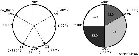
| Normal axis | -30 to +90 degrees |
| Left axis deviation | -30 to -90 degrees |
| Right axis deviation | +90 to +/-180 degrees |
| Indeterminate (extreme) axis deviation | -90 to +/-180 degrees |
Since lead I and aVF are perpendicular to each other, you can use those two leads to quickly determine axis.
Lead I runs from right to left across a patient's body, positive at the left hand.
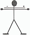
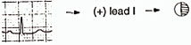
If the QRS in lead I is positive (mainly above the baseline), the direction of depolarization will be in the positive half (right half) of the circle above. You can make a diagram and shade in the positive half of the circle.
Lead aVF runs from top to bottom across a patient's body, positive at the feet.
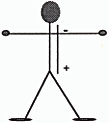
If the QRS in lead aVF is positive (mainly above the baseline), the direction of depolarization will be in the positive half (lower half) of the circle above. You can make a diagram and shade in the positive half of the circle:
![]()
To find the axis, overlap the two circles.
The common shaded area is the quadrant in which the axis lies.
In this
example, the axis lies in the normal quadrant, which on a patient,
points down and to the left.
![]()
You can repeat this process for any two
leads, but I and aVF are the classic places to look.
If you realize
that there are two leads to consider and a positive (+) or (-) orientation
for each lead, there would be four possible combinations.
Memorize the following axis guidelines:
Lead
I
|
Lead
aVF
|
|
| 1. Normal axis (0 to +90 degrees) | Positive
|
Positive
|
| 2. Left
axis deviation (-30 to -90). Also check lead II. To be true left axis deviation, it should also be down in lead II. If the QRS is upright in II, the axis is still normal (0 to -30) |
Positive
|
Negative
|
| 3. Right axis deviation (+90 to +180) | Negative
|
Positive
|
| 4. Indeterminate axis (-90 to -180) | Negative
|
Negative
|
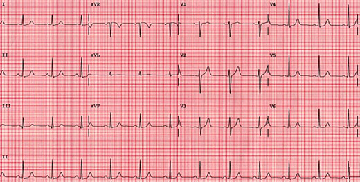
Normal axis
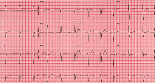
Left axis deviation
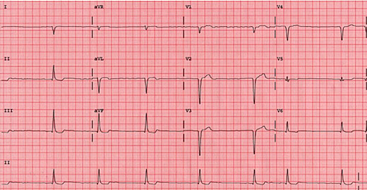
Right axis deviation
The bottom line is, if the axis is shifted out of the normal quadrant, evaluate the reasons for this.
Differential Diagnosis |
|
| Left axis deviation | LVH, left anterior fascicular block, inferior wall MI |
| Right axis deviation | RVH, left posterior fascicular block, lateral wall MI |
[ ECG index ]